Introduction: The Criticality of a Perfect Fit
Digital dentistry, powered by intraoral scanners (IOS), promises precision and efficiency. However, the reality often presents challenges: digital impressions that don’t fit the final lab work. This gap between digital promise and clinical reality leads to significant complications like open margins, occlusal discrepancies, and costly remakes. Studies show that while IOS are accurate, operator technique and data processing errors are often the root cause, not the hardware itself. Even minor discrepancies (e.g., 50 μm) can increase risks like secondary caries
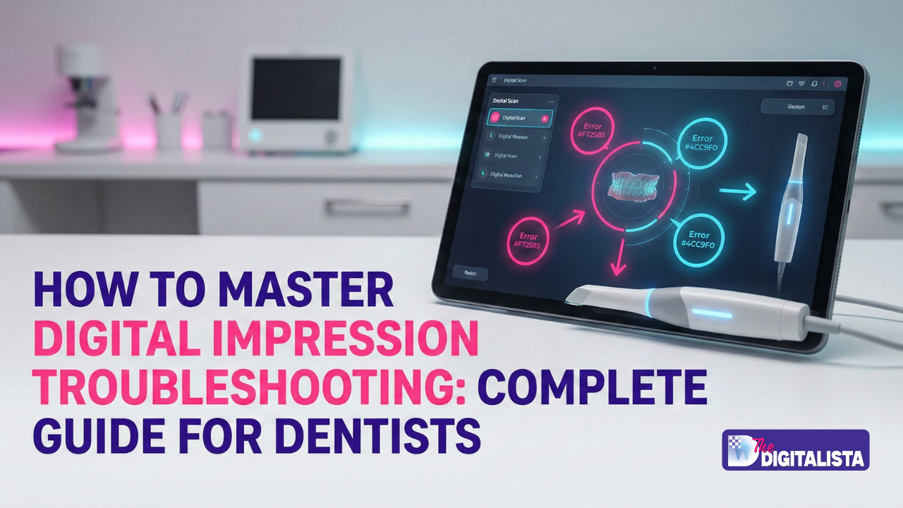
This guide provides a clinical roadmap to mastering the digital impression workflow. We will explore key technical problem areas, offer step-by-step solutions, and share advanced optimization techniques to ensure predictable, high-quality outcomes.
Section 1: Problem Overview & Clinical Context
Establishing Authority: Why Accuracy is Non-Negotiable
For the digital clinician, the accuracy of the digital impression is the foundation upon which all subsequent restorative work is built. This is not simply about capturing the shape of the teeth; it’s about translating complex, three- dimensional biological information—marginal integrity, emergence profile, tissue height, and occlusal scheme—into a mathematically precise digital file (typically an STL, PLY, or OBJ).
Clinical data consistently validates the high potential of digital scanning. A 2024 systematic review confirmed that the best IOS devices achieve full-arch accuracy within 40 μm, comparable to or better than PVS impressions, provided the correct technique is employed. However, this precision is fragile.
The clinical consequence of an ill-fitting restoration is severe:
– Financial Impact: Remakes due to fit issues can cost a practice thousands annually in lab fees, material waste, and lost chair time. A single remake erodes the profitability of the original procedure.
– Patient Trust: Repeated chairside adjustments or, worse, having to re-impress, damages patient confidence, and affects the perceived quality of your digital practice.
– Clinical Integrity: Open margins or over-contoured restorations increase plaque retention, accelerate periodontal breakdown, and mandate costly future interventions. A misfit is a clinical failure waiting to happen.
In the digital era, accountability is shared. When a restoration doesn’t fit, the potential causes range from inadequate preparation (clinical issue) to poor scanning protocol (operator issue), to file distortion (technical issue), to inaccurate milling/printing (lab issue).
Mastering troubleshooting means being able to pinpoint the exact weak link in the chain.

Section 2: Key Problem Areas in Digital Impression Capture & Transfer
The journey from the patient’s mouth to the milled crown involves multiple steps, each a potential point of failure. When a digital impression doesn’t fit the lab work, the cause usually falls into one of these key areas.
2.1. Soft Tissue Management and Prep Margin Capture
This is the single most common clinical mistake masquerading as a technical failure. The scanner can only capture what it can “see.”
- Cause: Blood, saliva, or unretracted gingiva obstructing the preparation margin. The scanner interpolates (guesses) data in areas it cannot capture clearly.
- Common Mistakes: Insufficient moisture control; using retraction cord that is too small or improperly placed; scanning immediately after removing the cord when fluid rebound is present.
- Clinical Impact: The lab design software receives an incomplete or fuzzy margin line, leading to an open margin on the final restoration. If the lab technician “guesses” the margin, the fit will be unpredictable

2.2. Scanner Movement and Stitching Errors (Registration Failure)
Intraoral scanners capture data in small video segments that are mathematically “stitched” or registered together to form the complete 3D model.
- Cause: Rapid scanner movement, scanning glossy or mirror-like surfaces (e.g., highly polished gold or wet enamel), or lack of unique anatomical reference points (e.g., scanning a long edentulous span).
- Why it Happens: The software loses tracking, resulting in data drift—a subtle, cumulative distortion that worsens the longer the scanning path. This is especially prevalent in full-arch scans.
- Clinical Impact: The resulting STL file is geometrically distorted. Crowns might fit an individual tooth but fail to align with the opposing arch or adjacent teeth, causing significant occlusal problems. This error is often described as a “stretched” or “warped” model.

2.3. Anti-Fogging and Calibration Protocol
All optical scanners are sensitive to temperature and humidity changes.
- Cause: Scanning while the mirror/lens is fogging up, or neglecting the manufacturer’s recommended calibration routine.
- Why it Happens: Fogging drastically reduces the amount of light reaching the sensor, leading to areas of data loss or “holes.” Calibration drift, which occurs over time and with temperature fluctuations, introduces a systematic (consistent) error in the distance measurement, affecting the scale of the entire model.
- Common Mistakes: Not waiting for the scanner tip to fully heat up in the mouth; skipping the weekly or bi-weekly calibration.
2.4. Post-Processing and File Compression Artifacts
- The impression data is often exported in a compressed file format for transfer to the lab.
- Cause: Using overly aggressive mesh decimation (reduction of the number of polygons) during the final export, or errors during the transfer.
- Clinical Impact: While decimation reduces file size (making transfer faster), it removes fine surface details, which include the critical anatomy of the margin line and the interproximal contacts. A file that is too small (e.g., under 15MB for a single quadrant) may lack the necessary resolution for precise lab design.
- Technical Specification: The industry standard for high-fidelity prosthetic work requires a mesh density that results in a file size typically above 40MB for a full arch. Always use the highest resolution export setting (e.g., binary STL) unless otherwise instructed.
Section 3: Step-by-Step Solutions for Guaranteed Fit
- Solving fit issues requires a systematic approach that addresses potential errors in the clinical, scanning, and transfer phases.
3.1. Clinical Protocol: Guaranteeing Margin Visibility
The highest resolution scanner cannot see through blood.
1. Hemostasis First: Achieve absolute hemostasis. Use an appropriate astringent (e.g., aluminum chloride) and a single or double-cord technique.
2. Air-Drying is Key: Thoroughly air-dry the entire preparation and adjacent tissue before scanning. Moisture interferes with optical capture.
3. Optimal Retraction: Ensure the gingival margin is retracted laterally and vertically. Remove the retraction cord just before scanning, or leave the smaller cord in place for a double-cord technique. The goal is a clear 360° view of the margin.
4. Inspect Visually: Use a mirror and magnification. If you can’t see the entire margin clearly, the scanner certainly can’t.
3.2. Scanning Protocol: Mastering the “Stitching” Process
Adhere strictly to a structured scanning sequence to prevent data drift.
1. Start Occlusally: Begin scanning the occlusal surface of the prepared tooth and the adjacent teeth to establish a strong registration base.
2. Follow the Sequence: Use a Z-pattern or manufacturer-recommended sequence. Transition from occlusal, roll lingually/palatally, sweep to the proximal, and roll buccally/facially, finishing back on the occlusal. Maintain slow, continuous movement.
3. Dedicated Margin Scan: Perform a slow, dedicated pass specifically targeting the margin line. Use the scanner’s magnification or “live color map” feature (if available) to ensure the margin is completely captured in high-density green or blue (indicating complete data capture).
4. Over-Scan: In the quadrant, ensure you have captured at least one full tooth anterior and posterior to the preparation. This gives the software sufficient, unique anatomy for registration.
5. Check for Holes: Review the final scan for “holes” (unscanned areas) or areas with fuzzy data. If present, re-scan that specific area with an insertion movement (placing the scanner tip directly over the hole and slowly pulling out) rather than a sweeping motion.
3.3. File Management and Laboratory Communication
Prevent technical transfer errors by establishing clear protocols.
1. Use High-Fidelity Export: Always export the scan in the highest resolution format. For STL, choose “binary” over “ASCII” if available, as it is a smaller file with the same fidelity. Do not use mesh decimation unless the lab explicitly requests a smaller file.
2. Clear File Naming: Include essential information for each case : PatientName _ToothNumber _PrepDate _ScannerModel.
3. Communicate Margin: Do not assume the lab can find the margin. If your scanner software allows, pre-mark the margin digitally before sending. This serves as a vital check and communication point.
4. Confirm Data: Send the lab a high-resolution screenshot of the prep in isolation, demonstrating the clear margin capture, alongside the full scan files.
Section 4: Best Practices & Pro Tips for Workflow Optimization
Moving beyond basic troubleshooting, these insider tips will help your practice
move from merely fixing problems to preventing them entirely.
4.1. The “Two-Step Scan Check”
Implement this simple, two-step quality assurance before sending any file:
1. The Marginal Check (Focus on Fit): Zoom in on the preparation margin. Rotate the model in 360 degrees. Look for any areas where the margin is faint, jagged, or where the scan data appears interpolated (smooth, unnatural-looking surface). If the margin disappears or appears doubled,
re-scan the area immediately.
2. The Occlusal Check (Focus on Stability): View the antagonist and working models together in the bite registration. Place a virtual “ruler” or measurement tool across the occlusal plane. Look for any discrepancies in the posterior segments, especially a “frown” or “smile” curve distortion in the arch, indicating a stitching error.
4.2. Workflow Optimization Pro Tips
- Dedicated Scanning Assistant: Train a dental assistant to be the “Scanning Master.” Standardizing the operator significantly reduces technique variability.
- Isolate and Retract: Use small, black triangle shields or contrast spray on highly glossy areas (like highly polished abutments or anterior incisal edges) to provide better texture for the optical tracking software.
- Know Your Scanner’s Limitations: Full-arch scans are more challenging with certain entry-level scanners. For large bridges or full arches, consider a reference marker technique or switch to a proven high-accuracy device.
Quick-Fix Cheat Sheet
| Issue | Cause | Chairside Fix |
|---|---|---|
| Holes | Fast move/wet | Tip-over-hole, pull slow |
| Bite Off | Few contacts | Re-scan 3+ points in CR |
| Too Big/Small | Calibration | Recalibrate now |

Section 5: Advanced Techniques & Software Comparison
For complex cases or persistent fit issues, advanced techniques and a clear
understanding of the design software’s role are essential.
Reference Body and Segmented Stitching
This technique is vital for managing distortion in long span bridges and full
arches.
| Technique | Description | Pros | Cons |
|---|---|---|---|
| Reference Body Stitch | Placing dedicated high-contrast markers (often small titanium spheres or custom abutments) in the arch before scanning. | Provides absolute, non-anatomical reference points; drastically reduces cumulative error. | Requires extra chair time for placement; initial cost for markers. |
| Segmented Scanning | Scanning the arch in 2-3 smaller segments and stitching them together outside the IOS software using a third- party program. | Excellent for high- distortion areas (e.g., large palatal vaults); better quality control. | More complex workflow; requires advanced design software knowledge. |
Advanced Hacks for Tough Cases
For full-arches, add scan bodies or segment (anterior/posterior). Tools like Blender fix meshes post-scan. Photogrammetry helps multi-implant arches over 4 units.
Your Path to Flawless Scans
Grab our free Digital Impression Checklist—Printable + Audio guide, game-changing.
Head to The Digitalista Checklist or DM “SCAN FIX” for a 15-min workflow audit. Track your next 10 scans; watch fits soar.
